
Imaging Center
Texas A&M College of Dentistry's Oral and Maxillofacial Imaging Center offers sophisticated diagnostic imaging services to health professionals. An integral part of a nationally recognized oral health educational institution, this full-service imaging facility specializes in assessing conditions of the head and neck region.
About the imaging center
The center can assist in the diagnostic workup of temporomandibular joint dysfunction, dental implant site assessment, orthodontics, craniofacial anomalies, salivary gland dysfunction, trauma and general pathology. There is no cost to the referring clinician for the services, as patients are charged directly at the time of consultation. The center is equipped to provide directly acquired computed tomography (CT scan), 2-D and 3-D reformatted CT, conventional film tomography, plain film radiography (intraoral, cephalometric, hand/wrist, skull, sinus), panoramic radiography (including hybrid maxillofacial and scanographic images), contrast radiology (sialography), and digital photography. Orthodontic tracings and diagnostic models are available, as is magnetic resonance imaging (MRI), which is offered through a cooperative agreement. All imaging studies are accompanied by a written interpretive report dictated by faculty who are diplomates of the American Board of Oral and Maxillofacial Radiology.
Referral Information
For information, referral forms or appointments call (214) 828-8479Referring doctors should complete a direct referral form, which is available upon request. Appointments may be arranged by telephoning the receptionist at the Oral and Maxillofacial Imaging Center. The center is located on the fifth floor, in the Clinic and Education Building of Texas A&M College of Dentistry.
Email codimagingcenter@tamu.eduTemporomandibular Joints
Temporomandibular Joint Evaluation
Imaging the TMJ area may be indicated when patients present with pain and tenderness in the TMJ, clicking or crepitus, locking or dislocation. These symptoms may be related to pathologic changes in the hard and/or soft tissue components of the TMJ. TMJ imaging also may be useful for documenting condylar position prior to or following splint therapy, crepitus, locking, or dislocation.
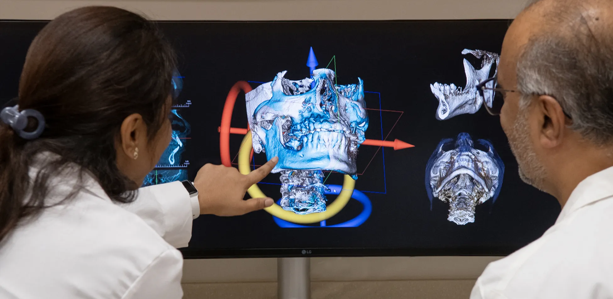
TMJ Computed Tomography
CT is the procedure of choice for studying the hard tissue components of the TMJ. Images acquired in one plane of view may be digitally enhanced and reconstructed by computer to show the condyle and glenoid fossa from virtually any perspective in both two and three dimensions.
Magnetic Resonance Imaging
MRI is a noninvasive procedure that does not use X-rays. It is the only method available that will directly and consistently show soft tissue components of the TMJ. MRI is useful for locating the position of the disk and for the assessment of inflammatory conditions of the TMJ area.
Dental Implants
Implant Site Assessment
Imaging of the residual alveolar ridge and adjacent structures is indicated both preoperatively and postoperatively in the placement of dental implants. In addition to ruling out pathology, imaging provides multidirectional views that allow the clinician to plan and follow the case for an optimal result.
Implant Computed Tomography
Axial images are reconstructed into multiple cross-sectional, panoramic and axial views. In addition to osseous morphology, the location of significant anatomical structures such as the mandibular and mental canals, maxillary sinus, nasal fossa and submandibular fossa are visualized for optimal implant placement.
Conventional Film Tomography
Conventional film tomography, like CT, provides cross-sectional images for studying the morphology of the intended implant site. The location of significant adjacent anatomical structures can be visualized with confidence. This technique is the procedure of choice for single implant sites.
Postoperative Implant Assessment
Intraoral and panoramic radiographs are usually adequate for postoperative implant assessment. Several views may be required for optimal visualization of the implant fixture. More complex cases may require conventional or computed tomography.
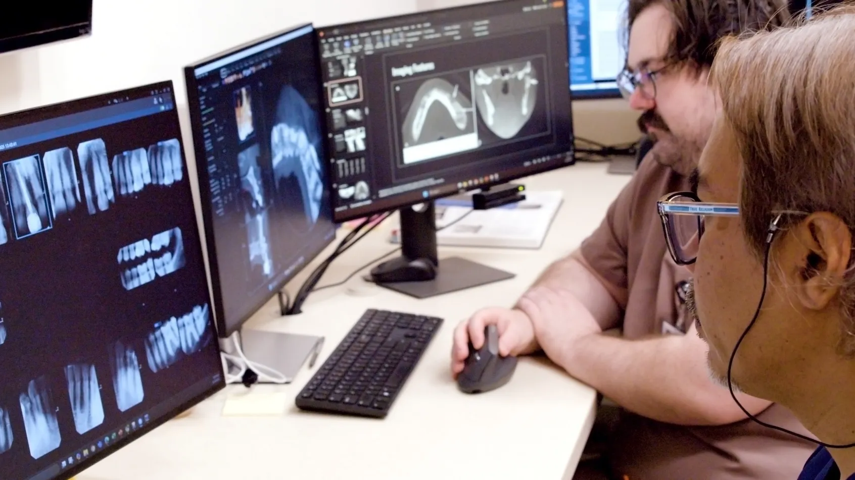
Cone Beam CT Interpretation Service
The Imaging Center offers interpretation of all imaging studies of the maxillofacial region acquired in your office through our teleradiology consult service via a secure server, for a nominal fee. Studies can be submitted under the “routine” or “rush” category based on the level of urgency. You will be assigned an account to upload your scans to the Imaging Center for prompt interpretation. Any further imaging recommendations as necessary are included in the reports which have an average turnaround time of 48-72 hours. All “rush” cases are sent out within 24 hours.